
Introduction to Leukemia Inhibitory Factor
Leukemia Inhibitory Factor, commonly abbreviated as LIF, is a multifunctional cytokine belonging to the interleukin-6 cytokine family. It was initially discovered because of its ability to induce differentiation and suppress proliferation in specific leukemic cell lines. However, subsequent research demonstrated that this molecule possesses a remarkably broad spectrum of biological activities extending far beyond hematological regulation.
During its early characterization, several independent laboratories identified proteins with apparently unrelated biological functions. Some factors inhibited embryonic stem cell differentiation, others stimulated proliferation of hematopoietic cells, triggered inflammatory responses in liver tissue, altered neuronal phenotype, or influenced lipid metabolism in adipocytes. Surprisingly, all these biological activities were later attributed to the same cytokine, now known as Leukemia Inhibitory Factor.
Today, LIF is recognized as one of the most pleiotropic cytokines in mammalian biology. It participates in stem cell maintenance, embryonic implantation, neural development, immune regulation, bone remodeling, muscle regeneration, endocrine signaling, cardiovascular protection, inflammatory responses, and cancer progression. Its versatility arises from the complexity of its receptor system and the diversity of intracellular signaling pathways activated in different cellular environments.
Molecular Structure of LIF
Biochemical Characteristics of the LIF Glycoprotein
LIF is synthesized as a precursor protein that undergoes post-translational processing before becoming biologically active. Mature LIF exists as a glycoprotein with a compact and highly stable three-dimensional structure typical of the interleukin-6 cytokine family.
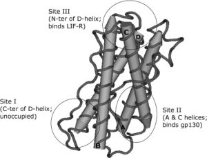
Structural studies using crystallography and nuclear magnetic resonance techniques revealed that LIF adopts a characteristic four-helix bundle architecture. This structural organization is stabilized by multiple disulfide bridges that maintain protein integrity and contribute to receptor binding efficiency.
One particularly important feature of the molecule is its exceptional stability. Experimental analyses demonstrated that LIF maintains structural rigidity even under physiologically stressful conditions. This stability likely contributes to its prolonged biological activity and efficient receptor engagement.
The N-terminal region of the molecule plays a major role in receptor interaction. Structural integrity within this domain is essential for high-affinity binding to receptor subunits and successful activation of downstream signaling cascades.
The LIF Receptor Complex
Composition of the LIF Receptor System
The biological activity of LIF depends on its interaction with a specialized receptor complex composed primarily of two signaling chains:
- gp130
- Leukemia Inhibitory Factor Receptor beta (LIFRβ)
Unlike several related cytokines that require additional alpha-chain receptors for signaling, LIF can activate intracellular signaling pathways through a simpler receptor assembly. This receptor organization distinguishes LIF from other members of the interleukin-6 cytokine family.
The receptor complex forms after LIF simultaneously binds both gp130 and LIFRβ. These receptor chains contain intracellular domains associated with Janus kinases, allowing rapid initiation of signal transduction once receptor activation occurs.
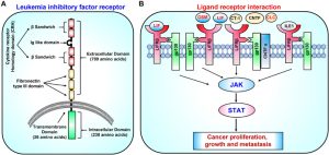
Structural Organization of LIFRβ
LIFRβ is a single-pass transmembrane protein characterized by a large extracellular domain containing multiple functional regions.
The extracellular portion includes cytokine-binding modules, immunoglobulin-like domains, and fibronectin type III domains. These structural elements coordinate ligand recognition and proper receptor alignment at the cell membrane.
The fibronectin domains act as molecular spacers that position intracellular signaling components appropriately for kinase activation. Once receptor dimerization occurs, associated kinases become activated through reciprocal phosphorylation events.
LIFRβ is not exclusive to LIF signaling. Several cytokines share this receptor chain, including:
- Oncostatin M
- Cardiotrophin-1
- Ciliary neurotrophic factor
- Cardiotrophin-like cytokine
This receptor sharing partially explains the overlapping biological functions observed among cytokine family members.
Mechanisms of LIF-Receptor Interaction
High-Affinity Binding and Signal Initiation
LIF displays exceptionally strong receptor-binding affinity. The cytokine independently interacts with both gp130 and LIFRβ, allowing rapid formation of a signaling-competent receptor complex.
Unlike interleukin-6, whose receptor assembly occurs through a highly cooperative and ordered process, LIF receptor binding appears less dependent on sequential assembly. The interaction is primarily driven by favorable thermodynamic forces and requires relatively limited structural rearrangement upon complex formation.
This efficient binding mechanism contributes to rapid signal initiation and sustained cellular responses. Experimental kinetic analyses demonstrated that receptor dissociation occurs very slowly, indicating prolonged signaling capacity after ligand engagement.
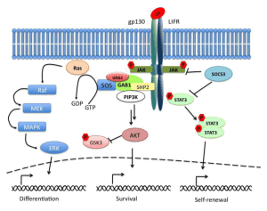
Intracellular Signaling Pathways Activated by LIF
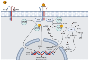
JAK/STAT Signaling Pathway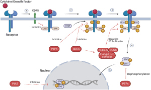
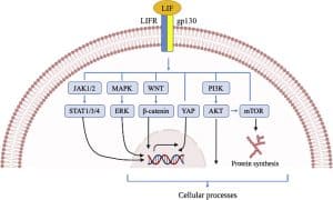
The most important signaling cascade activated by LIF involves the Janus kinase and Signal Transducer and Activator of Transcription pathway.
Unlike receptor tyrosine kinases, the LIF receptor itself lacks intrinsic enzymatic activity. Instead, intracellular receptor domains are constitutively associated with Janus kinases, particularly JAK1.
Upon ligand binding:
- Receptor-associated JAK molecules become activated
- Receptor intracellular domains undergo phosphorylation
- STAT proteins dock onto phosphorylated receptor sites
- STAT proteins become phosphorylated and activated
- Activated STAT dimers translocate to the nucleus
- Cytokine-responsive genes are transcribed
Among STAT family members, STAT3 is considered the dominant mediator of LIF biological activity.
STAT3 as a Master Regulator
STAT3 functions as a central transcriptional regulator controlling stem cell maintenance, survival, differentiation, and inflammatory responses.
Once activated, STAT3 promotes expression of genes associated with:
- Self-renewal
- Cell survival
- Pluripotency
- Metabolic regulation
- Anti-apoptotic activity
STAT3 also induces expression of suppressor proteins such as SOCS3, establishing a negative feedback loop that prevents excessive signaling.
This autoregulatory mechanism is essential because uncontrolled LIF signaling can produce severe developmental abnormalities and pathological tissue remodeling.
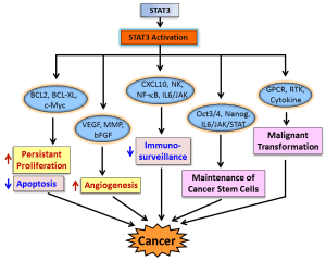
PI3K/AKT Signaling Pathway
LIF additionally activates the phosphoinositide 3-kinase pathway, an essential regulator of metabolism, cell survival, and proliferation.
Activation of PI3K leads to downstream stimulation of AKT, a kinase controlling numerous cellular processes including:
- Protein synthesis
- Cell-cycle progression
- Glucose metabolism
- Anti-apoptotic signaling
- Stem cell maintenance
In embryonic stem cells, PI3K/AKT signaling strongly supports pluripotency and self-renewal.
Experimental studies demonstrated that artificial activation of AKT signaling can maintain stem cell characteristics even in the absence of LIF, highlighting the importance of this pathway.
MAPK Signaling Cascade
LIF also stimulates the mitogen-activated protein kinase pathway through recruitment of adaptor proteins and phosphatases such as SHP2.
MAPK signaling generally promotes:
- Differentiation
- Cell proliferation
- Mitogenic responses
- Developmental progression
Interestingly, in embryonic stem cells, excessive MAPK activation favors differentiation rather than pluripotency. Therefore, stem cell maintenance depends on a delicate balance between STAT3/PI3K signaling and MAPK activity.
This illustrates how the same cytokine can trigger competing cellular programs depending on signaling intensity and cellular context.
LIF and Embryonic Stem Cells
Maintenance of Pluripotency
One of the most extensively studied functions of LIF is its role in maintaining mouse embryonic stem cells in an undifferentiated state.
Mouse embryonic stem cells require continuous LIF exposure to preserve:
- Self-renewal
- Pluripotency
- Long-term proliferation
- Resistance to differentiation
LIF achieves this primarily through STAT3 activation, although PI3K signaling also contributes significantly.
In contrast, human embryonic stem cells depend more heavily on fibroblast growth factor and activin signaling pathways, illustrating species-specific differences in pluripotency regulation.
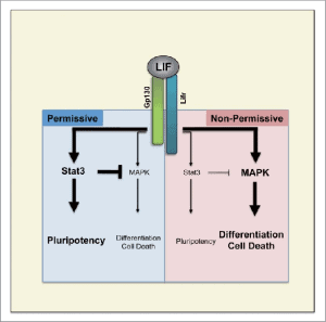
Role in Induced Pluripotent Stem Cells
LIF signaling also contributes to the generation and maintenance of induced pluripotent stem cells.
During cellular reprogramming, transcription factors associated with pluripotency become activated in coordination with LIF-responsive signaling pathways. These interactions facilitate the transition from differentiated somatic cells toward pluripotent stem cell states.
Consequently, LIF has become an essential component of many stem cell culture systems used in regenerative medicine and developmental biology research.
LIF in Reproductive Biology
Embryo Implantation and Fertility
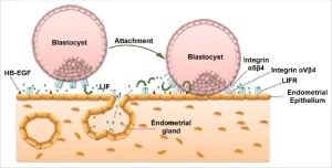
LIF plays a crucial role during embryo implantation and uterine receptivity.
During early pregnancy, the endometrium produces elevated levels of LIF in preparation for blastocyst implantation. Both the embryo and maternal endometrial tissues express functional LIF receptors.
Experimental evidence demonstrated that female mice lacking LIF are infertile because embryos fail to implant properly. Importantly, the defect originates from the maternal environment rather than the embryo itself.
Reduced uterine LIF expression has also been associated with infertility in humans, suggesting that impaired LIF signaling may contribute to implantation failure and reproductive disorders.
Placental Development
LIF influences trophoblast differentiation and placental formation.
Proper regulation of trophoblast invasion and placental architecture requires tightly controlled LIF signaling. Excessive signaling can disrupt placental organization, whereas insufficient activity impairs implantation and fetal support.
These findings highlight the importance of balanced cytokine signaling during pregnancy.
LIF in Bone Biology
Bone Remodeling and Osteoblast Differentiation
LIF participates in skeletal homeostasis by influencing osteoblast differentiation and bone remodeling.
Osteoblasts express functional LIF receptors and respond to LIF stimulation by promoting bone-forming activity. LIF also suppresses adipogenic differentiation within bone marrow stromal populations.
Additionally, LIF regulates expression of molecules involved in osteoclast activation and communication between bone-forming and bone-resorbing cells.
Loss-of-function mutations affecting the LIF receptor have been associated with severe skeletal abnormalities characterized by impaired bone development and structural deformities.
Neurological Functions of LIF
Neural Development and Injury Response
LIF exerts extensive effects throughout the nervous system.
Originally identified as a factor capable of altering neurotransmitter expression in sympathetic neurons, LIF is now known to regulate:
- Neural differentiation
- Neuron survival
- Axonal regeneration
- Astrocyte development
- Oligodendrocyte maturation
- Myelination processes
Importantly, many neural functions of LIF become especially prominent following tissue injury.
After nerve damage, LIF expression increases dramatically in injured tissues. Retrograde transport mechanisms deliver LIF signals back to neuronal cell bodies, promoting survival and regenerative responses.
Glial Cell Regulation
LIF strongly influences glial cell populations, including astrocytes and oligodendrocytes.
In the central nervous system, LIF supports:
- Astrocyte differentiation
- Oligodendrocyte maturation
- Myelin repair
- Neural stem cell maintenance
Experimental models demonstrated improved remyelination following LIF administration, suggesting potential applications in demyelinating disorders.
LIF and Muscle Regeneration
Skeletal Muscle Repair
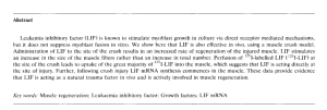
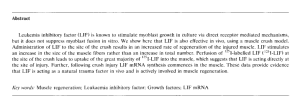
LIF expression increases substantially following muscle injury and physical exercise.
In skeletal muscle tissue, LIF promotes:
- Myoblast proliferation
- Muscle regeneration
- Protection against atrophy
- Neuromuscular repair
Although developmental muscle formation appears relatively normal in LIF-deficient animals, regenerative responses after injury become significantly impaired.
This suggests that LIF functions primarily as an injury-response cytokine rather than a developmental determinant in muscular tissues.
Cardiovascular Functions of LIF
LIF also exerts protective effects within cardiac tissue.
Experimental studies demonstrated that LIF can:
- Enhance cardiomyocyte survival
- Reduce ischemia-reperfusion injury
- Stimulate protective antioxidant pathways
- Improve recovery after myocardial infarction
Some cardioprotective effects involve activation of PI3K/AKT survival signaling and induction of stress-response genes.
These findings generated interest in potential therapeutic applications for cardiovascular disease and tissue repair.
Immunological and Hematopoietic Functions
Immune Regulation
Although initially discovered in leukemic cells, LIF displays relatively modest direct effects on hematopoietic populations compared with other cytokines.
However, LIF exerts important immunomodulatory activities by influencing:
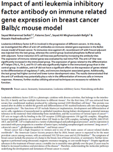
- Regulatory T-cell differentiation
- Inflammatory cytokine production
- Acute phase responses
- Immune tolerance
LIF promotes development of regulatory T cells while suppressing pro-inflammatory immune responses, suggesting an anti-inflammatory and tolerogenic role.
These activities may contribute to maternal-fetal immune tolerance during pregnancy and protection against excessive inflammatory injury.
LIF and Cancer Biology
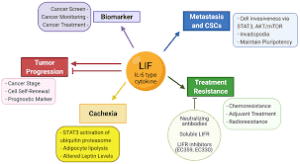
Tumor Progression and Therapy Resistance
Abnormal LIF expression has been detected in multiple human cancers, including:
- Breast cancer
- Colorectal cancer
- Skin cancer
- Nasopharyngeal carcinoma
In many tumors, LIF promotes:
- Tumor growth
- Metastatic behavior
- Survival signaling
- Resistance to chemotherapy
- Resistance to radiotherapy
LIF-mediated activation of STAT3 and other pathways can suppress tumor suppressor functions while enhancing survival mechanisms.
Tumor hypoxia has also been shown to stimulate LIF expression, further linking this cytokine to aggressive cancer phenotypes.
Clinical Perspectives and Therapeutic Potential
Because of its broad biological functions, LIF has attracted considerable interest for therapeutic applications in:
- Infertility treatment
- Stem cell culture systems
- Neural repair
- Muscle regeneration
- Cardiovascular protection
- Immunomodulation
However, clinical translation remains challenging due to the complexity and context-dependent nature of LIF signaling.
Excessive or inappropriate activation may produce undesirable effects including abnormal tissue remodeling, inflammation, fibrosis, or tumor progression.
Future therapeutic strategies will likely require highly targeted and temporally controlled modulation of LIF pathways.
Conclusion
Leukemia Inhibitory Factor is one of the most biologically versatile cytokines identified in mammalian systems. Initially recognized for its ability to suppress leukemic cell proliferation, LIF is now understood to regulate a vast network of physiological processes including stem cell maintenance, embryonic implantation, neural survival, bone remodeling, immune tolerance, muscle regeneration, and tissue repair.
Its biological diversity arises from sophisticated receptor interactions and activation of multiple intracellular signaling pathways, particularly the JAK/STAT3, PI3K/AKT, and MAPK cascades.
One of the most fascinating aspects of LIF biology is its context-dependent behavior. In some cellular systems LIF promotes differentiation, whereas in others it preserves pluripotency and self-renewal. Similarly, it can stimulate proliferation in certain tissues while inducing growth arrest or apoptosis in others.
These seemingly contradictory activities emphasize the importance of cellular context, receptor composition, chromatin organization, and signaling balance in determining biological outcomes.
Despite decades of research and numerous experimental studies, many aspects of LIF biology remain incompletely understood. Nevertheless, LIF continues to represent a major focus in stem cell biology, regenerative medicine, developmental biology, immunology, and cancer research.